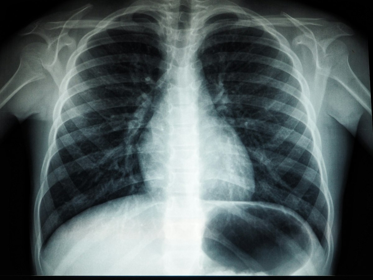
Few areas of health technology attract bolder claims than AI for medical images. Headlines suggest software that reads scans better than doctors. The reality is more specific and more interesting. Some uses are well established and cleared for use. Others look strong in a paper and weaken in a hospital.
This is a technology and operations discussion, not clinical advice. If you run or buy imaging systems, the question is how to judge claims carefully, and the answer is mostly about evidence and workflow.
Why imaging suits machine learning
Medical images are structured data. A chest X-ray, a retinal photograph or a CT slice is a grid of values, and pattern recognition on grids is what modern machine learning does well. There is also a long record of digitised, labelled images stored in standard formats such as DICOM, which gives researchers material to train on.
That explains the early progress. It does not explain whether a given tool helps patients when deployed. Those are different questions, and confusing them is the root of most overstated claims.
What is reasonably established
Regulators in several regions, including the US FDA and bodies in the EU and UK, have cleared a number of AI based imaging products. These are mostly narrow tools doing a defined task. Typical examples include:
- Flagging studies that may contain an urgent finding so they are read sooner, which is a prioritisation function.
- Highlighting regions of an image for a clinician to review, as a second pair of eyes.
- Measuring and quantifying structures consistently, such as sizing a finding or calculating a volume.
- Assisting with screening programmes in specific, well studied settings.
A clearance means a regulator reviewed evidence for a stated intended use. It does not mean the tool is right for every patient population, scanner or hospital. Read the intended use statement. It is usually narrower than the marketing.
What is less certain
Several gaps recur, and they deserve scepticism.
Performance on different populations and equipment. A model trained on images from a few hospitals can lose accuracy when it meets different scanners, protocols or patient demographics. This is often called a generalisation problem. A strong result at the developer's site is a starting point, not proof for yours.
Retrospective versus real world results. Many studies test a model on past, curated images. Real clinics bring poor quality scans, unusual cases and time pressure. Prospective studies that follow outcomes in live practice are harder, slower and rarer, and they matter more.
Accuracy versus benefit. A tool can detect a finding accurately and still fail to improve care. Finding more small abnormalities may lead to more follow up tests without changing outcomes. Evidence on patient benefit, not only detection rates, is what to look for.
Human and machine together. How a radiologist responds to an AI suggestion is a research subject in itself. Automation bias, where people over trust a prompt, and the reverse, where they ignore it, both appear. The combined system's performance is what counts.
Drift over time. Equipment gets upgraded, protocols change and disease patterns shift. A model that performed well at purchase can degrade quietly without monitoring.
Questions to ask a vendor
If you are evaluating a product, a plain set of questions cuts through a great deal.
- What exact task and population is it cleared or validated for?
- Was it tested on data from sites unlike the ones it was built on, and was that data kept separate from training?
- Is there evidence from live clinical use, and has it been published or independently reviewed?
- How does it perform for subgroups, and where does it do worst?
- What happens on the cases it gets wrong, and how are those surfaced?
- How is performance monitored after installation?
A serious vendor will answer these directly and admit limits. Evasion is information too.
The operational side
Much of the real work is not the model. It is integration. Images flow from scanners through a PACS, results must return to the reporting workflow, and the output must appear where the clinician is already working. A tool that needs a separate login and a separate screen tends to be ignored.
Plan for standards based integration with existing systems rather than bespoke connections. Make the interface show clearly what the AI did and did not assess, so no one assumes more than was done. Keep an audit trail recording which version of the model produced which output, because a model update changes behaviour and you will want to know which version read which scan. Give clinicians a route to report errors, and make sure someone acts on those reports.
Data governance needs its own attention: where images are processed, what leaves the hospital, and on what legal basis. Imaging data is sensitive health data under laws such as the Nigeria Data Protection Act 2023 and GDPR, and a vendor that processes it in the cloud is a processor you must be able to account for.
Staffing matters too. Someone has to own the tool after the launch meeting: reviewing the error reports, talking to the vendor about updates, and deciding when to switch it off. Without a named owner, even a good tool decays into an ignored banner on a screen.
Context in lower resource settings
Imaging AI is sometimes proposed as a way to extend scarce specialist capacity, where radiologists are few and distances are long. The motivation is understandable. The cautions apply with extra force: image quality varies, equipment may differ from what the model learned on, and follow up care must exist for anything the tool flags. A finding with no pathway to treatment is not an improvement. Local validation matters more, not less, and so does training for the staff who will see the output.
A measured approach
Caution here is not hostility to the technology. It is how useful tools earn trust.
Start with a narrow, well defined use such as worklist prioritisation, where the AI changes order rather than diagnosis. Run it in shadow mode first, where it produces outputs that are compared to clinicians' decisions without influencing them. Review the results with the clinicians who will use it. Then expand only as the local evidence supports it.
The honest summary is that AI in imaging is real, useful in specific roles and often unproven beyond them. Treat each claim as a question about a particular task, population and workflow, and ask for the evidence that fits your own.